
ਤਸਵੀਰ:RestingStateModels.jpg

ਇਸ ਝਾਤ ਦਾ ਅਕਾਰ: 563 × 599 ਪਿਕਸਲ. ਹੋਰ ਰੈਜ਼ੋਲਿਊਸ਼ਨਜ਼: 226 × 240 ਪਿਕਸਲ | 451 × 480 ਪਿਕਸਲ | 796 × 847 ਪਿਕਸਲ.
ਅਸਲ ਫ਼ਾਈਲ (796 × 847 ਪਿਕਸਲ, ਫ਼ਾਈਲ ਅਕਾਰ: 158 KB, MIME ਕਿਸਮ: image/jpeg)
ਸਾਰ
| ਵੇਰਵਾ |
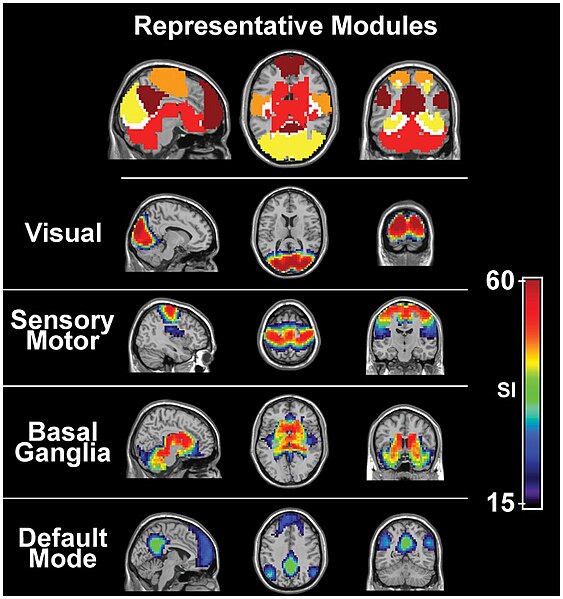
English: Study showing four functional networks that were found to be highly consistent across subjects. These modules include the visual (yellow), sensory/motor (orange) and basal ganglia (red) cortices as well as the default mode network (precuneus/posterior cingulate, inferior parietal lobes, and medial frontal gyrus; maroon). Overlap among these modules was present but minimal (white).[1] |
| ਮਿਤੀ | |
| ਸਰੋਤ |
PLoS One. 2012; 7(8): e44428. Published online 2012 August 31 |
| ਲਿਖਾਰੀ | Malaak N. Moussa, Matthew R. Steen, Paul J. Laurienti, and Satoru Hayasaka |
| Other versions |
 |
ਲਸੰਸ
This file is licensed under the Creative Commons Attribution 2.5 Generic license.
- ਤੁਹਾਨੂੰ ਖੁੱਲ੍ਹ ਹੈ:
- ਸਾਂਝਾ ਕਰਨ ਦੀ – ਰਚਨਾ ਨੂੰ ਕਾਪੀ, ਵੰਡਣਾ ਅਤੇ ਭੇਜਣਾ
- ਮੁੜ-ਰਲ਼ਾਉਣ ਦੀ – ਰਚਨਾ ਨੂੰ ਢਾਲਣਾ
- ਥੱਲੇ ਲਿਖੀਆਂ ਸ਼ਰਤਾਂ ਹੇਠ:
- ਗੁਣਾਂ ਦੀ ਦੱਸ – ਉਚਿਤ ਕ੍ਰੈਡਿਟ ਦੇਵੋ, ਲਾਇਸੰਸ ਦਾ ਇੱਕ ਲਿੰਕ ਪ੍ਰਦਾਨ ਕਰੋ ਅਤੇ ਇਹ ਦਰਸਾਓ ਕਿ ਕੀ ਤਬਦੀਲੀਆਂ ਕੀਤੀਆਂ ਗਈਆਂ ਸਨ। ਤੁਸੀਂ ਇਹ ਕਿਸੇ ਵੀ ਵਾਜਬ ਤਰੀਕੇ ਨਾਲ ਕਰ ਸਕਦੇ ਹੋ, ਪਰ ਇਹ ਤਰੀਕਾ ਅਜਿਹਾ ਨਹੀਂ ਹੋਣਾ ਚਾਹੀਦਾ ਜੋ ਇਹ ਦਰਸਾਵੇ ਕਿ ਲਾਇਸੰਸਕਰਤਾ ਤੁਹਾਨੂੰ ਜਾਂ ਤੁਹਾਡੀ ਵਰਤੋਂ ਦਾ ਸਮਰਥਨ ਕਰਦਾ ਹੈ।
- ↑ Moussa, Malaak N. (2012-08-31). "Consistency of Network Modules in Resting-State fMRI Connectome Data". PLoS ONE 7 (8): e44428. Public Library of Science (PLoS). DOI:10.1371/journal.pone.0044428. ISSN 1932-6203.
ਫ਼ਾਈਲ ਦਾ ਅਤੀਤ
ਤਾਰੀਖ/ਸਮੇਂ ’ਤੇ ਕਲਿੱਕ ਕਰੋ ਤਾਂ ਉਸ ਸਮੇਂ ਦੀ ਫਾਈਲ ਪੇਸ਼ ਹੋ ਜਾਵੇਗੀ।
| ਮਿਤੀ/ਸਮਾਂ | ਨਮੂਨਾ | ਨਾਪ | ਵਰਤੋਂਕਾਰ | ਟਿੱਪਣੀ | |
|---|---|---|---|---|---|
| ਮੌਜੂਦਾ | 01:11, 28 ਨਵੰਬਰ 2012 | | 796 × 847 (158 KB) | Amorrissette3 | User created page with UploadWizard |
ਫ਼ਾਈਲ ਦੀ ਵਰਤੋਂ
ਇਹ ਫਾਈਲ ਹੇਠਾਂ ਦਿੱਤਾ ਸਫ਼ਾ ਵਰਤਦਾ ਹੈ:
ਫ਼ਾਈਲ ਦੀ ਵਿਆਪਕ ਵਰਤੋਂ
ਇਸ ਫ਼ਾਈਲ ਨੂੰ ਹੋਰ ਹੇਠ ਲਿਖੇ ਵਿਕੀ ਵਰਤਦੇ ਹਨ:
- as.wikipedia.org ਉੱਤੇ ਵਰਤੋਂ
- da.wikipedia.org ਉੱਤੇ ਵਰਤੋਂ
- de.wikipedia.org ਉੱਤੇ ਵਰਤੋਂ
- en.wikipedia.org ਉੱਤੇ ਵਰਤੋਂ
- es.wikipedia.org ਉੱਤੇ ਵਰਤੋਂ
- fa.wikipedia.org ਉੱਤੇ ਵਰਤੋਂ
- hu.wikipedia.org ਉੱਤੇ ਵਰਤੋਂ
- ml.wikipedia.org ਉੱਤੇ ਵਰਤੋਂ
- pt.wikipedia.org ਉੱਤੇ ਵਰਤੋਂ
- sr.wikipedia.org ਉੱਤੇ ਵਰਤੋਂ
- th.wikipedia.org ਉੱਤੇ ਵਰਤੋਂ
- tr.wikipedia.org ਉੱਤੇ ਵਰਤੋਂ
- www.wikidata.org ਉੱਤੇ ਵਰਤੋਂ
- zh.wikipedia.org ਉੱਤੇ ਵਰਤੋਂ