ਅੰਡਕੋਸ਼ ਕੈਂਸਰ
| ਅੰਡਕੋਸ਼ ਕੈਂਸਰ | |
|---|---|
 | |
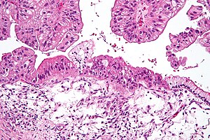
| ਮਾਈਕਰੋਗ੍ਰਾਫ਼ | |
| ਵਿਸ਼ਸਤਾ | ਓਨਕੋਲੋਜੀ, ਗਾਇਨਕੋਲੋਜੀ |
| ਲੱਛਣ | Early: vague[1] Later: bloating, pelvic pain, abdominal swelling, loss of appetite[1] |
| ਆਮ ਸ਼ੁਰੂਆਤ | Usual age of diagnosis 63 years old[2] |
| ਕਿਸਮ | Ovarian carcinoma, germ cell tumor, sex cord stromal tumor[3] |
| ਜ਼ੋਖਮ ਕਾਰਕ | Never having children, hormone therapy after menopause, fertility medication, obesity, genetics[3][4][5] |
| ਜਾਂਚ ਕਰਨ ਦਾ ਤਰੀਕਾ | Tissue biopsy[1] |
| ਇਲਾਜ | Surgery, radiation therapy, chemotherapy[1] |
| Prognosis | Five-year survival rate c. 45% (US)[6] |
| ਅਵਿਰਤੀ | 1.2 million (2015)[7] |
| ਮੌਤਾਂ | 161,100 (2015)[8] |
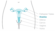
ਅੰਡਕੋਸ਼ ਕੈਂਸਰ, ਇੱਕ ਕੇਂਸਰ ਹੈ, ਜੋ ਅੰਡਕੋਸ਼ ਵਿੱਚ ਜਾਂ ਇਸ ਦੇ ਅੰਦਰ ਹੁੰਦਾ ਹੈ।[9] ਇਹ ਅਸਾਧਾਰਨ ਕੋਸ਼ਾਣੂ ਵਿੱਚ ਹੁੰਦਾ ਹੈ ਜਿਸ ਵਿੱਚ ਸ਼ੱਕ ਕਰਨ ਜਾਂ ਸਰੀਰ ਦੇ ਹੋਰ ਹਿੱਸਿਆਂ ਵਿੱਚ ਫੈਲਣ ਦੀ ਸਮਰੱਥਾ ਹੁੰਦੀ ਹੈ।[10] ਜਦੋਂ ਇਹ ਪ੍ਰਕਿਰਿਆ ਸ਼ੁਰੂ ਹੁੰਦੀ ਹੈ, ਕੋਈ ਵੀ ਜਾਂ ਸਿਰਫ ਅਸਪਸ਼ਟ ਲੱਛਣ ਹੋ ਸਕਦੇ ਹਨ। ਜਿਵੇਂ ਜਿਵੇਂ ਕੈਂਸਰ ਵਧਦਾ ਹੈ ਲੱਛਣ ਹੋਰ ਵੱਧ ਧਿਆਨ ਦੇਣ ਯੋਗ ਬਣਦੇ ਹਨ।[11] ਇਹਨਾਂ ਲੱਛਣਾਂ ਵਿੱਚ ਬਲੋਟਿੰਗ, ਪੇਲਵਿਕ ਦਰਦ, ਪੇਟ ਦੀਆਂ ਸੋਜ, ਅਤੇ ਭੁੱਖ ਦੇ ਨੁਕਸਾਨ ਸ਼ਾਮਲ ਹੋ ਸਕਦੇ ਹਨ। ਆਮ ਖੇਤਰ ਜਿਨ੍ਹਾਂ ਨਾਲ ਕੈਂਸਰ ਫੈਲ ਸਕਦਾ ਹੈ ਉਨ੍ਹਾਂ ਵਿੱਚ ਪੇਟ, ਲਿੰਫ ਗੁੱਛੇ, ਫੇਫੜੇ ਅਤੇ ਜਿਗਰ ਦੀ ਲਾਈਨਾਂ ਸ਼ਾਮਲ ਹਨ।[12]
ਅੰਡਕੋਸ਼ ਕੈਂਸਰ ਦਾ ਜੋਖਮ ਉਹਨਾਂ ਔਰਤਾਂ ਵਿੱਚ ਵੱਧ ਜਾਂਦਾ ਹੈ ਜਿਨ੍ਹਾਂ ਨੇ ਆਪਣੇ ਜੀਵਨ ਕਾਲ ਤੋਂ ਵੱਧ ਓਵੂਲੇਸ਼ਨ ਕੀਤਾ ਹੋਇਆ ਹੈ। ਇਸ ਵਿੱਚ ਉਹ ਲੋਕ ਵੀ ਸ਼ਾਮਲ ਹੁੰਦੇ ਹਨ ਜਿਨ੍ਹਾਂ ਦੇ ਕਦੇ ਬੱਚੇ ਨਹੀਂ ਸਨ, ਉਹ ਜਿਹੜੇ ਇੱਕ ਛੋਟੀ ਉਮਰ ਵਿੱਚ ਓਵੂਲੇਸ਼ਨ ਸ਼ੁਰੂ ਕਰਦੇ ਹਨ ਅਤੇ ਜਿਹੜੇ ਉਮਰ ਦੇ ਸਮੇਂ ਮੇਨੋਪੌਜ਼ ਵਿੱਚ ਪਹੁੰਚਦੇ ਹਨ। ਮਾਹਵਾਰੀ ਰੁਕਣਾ, ਉਪਜਾਊ ਦਵਾਈ ਅਤੇ ਮੋਟਾਪੇ ਤੋਂ ਬਾਅਦ ਹੋਰ ਖਤਰੇ ਦੇ ਕਾਰਕਾਂ ਵਿੱਚ ਹਾਰਮੋਨ ਥੈਰੇਪੀ ਸ਼ਾਮਲ ਹੈ।[4][5] ਜੋ ਖਤਰੇ ਨੂੰ ਘਟਾਉਂਦੇ ਹਨ ਉਹ ਹਾਰਮੋਨਲ ਜਨਮ ਨਿਯੰਤਰਣ, ਟਿਊਬਲ ਲਿਊਗੇਸ਼ਨ, ਅਤੇ ਦੁੱਧ ਚੁੰਘਾਉਣਾ ਹਨ। ਲਗਭਗ 10% ਮਾਮਲੇ ਵਿਰਾਸਤ ਵਾਲੇ ਜੈਨੇਟਿਕ ਜੋਖਮ ਨਾਲ ਸਬੰਧਤ ਹਨ; ਬੀ.ਆਰ.ਸੀ.ਏ.1 ਜਾਂ ਬੀ.ਆਰ.ਸੀ.ਏ.2 ਜੀਨਾਂ ਵਿੱਚ ਤਬਦੀਲੀਆਂ ਵਾਲੇ ਔਰਤਾਂ ਵਿੱਚ ਬਿਮਾਰੀ ਦੇ ਵਿਕਾਸ ਦੀ 50% ਸੰਭਾਵਨਾ ਹੈ। ਅੰਡਕੋਸ਼ ਕੈਂਸਰ ਦੀ ਸਭ ਤੋਂ ਆਮ ਕਿਸਮ, ਜਿਸ ਵਿੱਚ 95% ਤੋਂ ਵੱਧ ਕੇਸ ਹੁੰਦੇ ਹਨ, ਅੰਡਕੋਸ਼ ਕੈਂਸਰਿਨੋਮਾ ਹੈ। ਅੰਡਕੋਸ਼ਕ ਕੈਂਸਰਿਨੋਮਾ ਦੀਆਂ ਪੰਜ ਮੁੱਖ ਉਪ-ਰਿਪੋਜ਼ੀਆਂ ਹੁੰਦੀਆਂ ਹਨ, ਜਿਨ੍ਹਾਂ ਵਿੱਚੋਂ ਉੱਚ ਪੱਧਰੀ ਸੌਰਸ ਕਾਰਸਿਨੋਮਾ ਬਹੁਤ ਆਮ ਹੁੰਦਾ ਹੈ। ਮੰਨਿਆ ਜਾਂਦਾ ਹੈ ਕਿ ਇਹ ਟਿਊਮਰ ਅੰਡਾਸ਼ਯਾਂ ਨੂੰ ਢਕਣ ਵਾਲੇ ਸੈੱਲਾਂ ਵਿੱਚ ਸ਼ੁਰੂਆਤ ਕਰਦੇ ਹਨ, ਹਾਲਾਂਕਿ ਕੁਝ ਫਾਲੋਪੀਅਨ ਟਿਊਬਾਂ 'ਤੇ ਬਣਦੇ ਹਨ।[13] ਅੰਡਕੋਸ਼ ਦੇ ਕੈਂਸਰ ਦੀਆਂ ਘੱਟ ਆਮ ਕਿਸਮਾਂ ਵਿੱਚ ਜਰਮ ਦੇ ਸੈੱਲ ਟਿਊਮਰ ਅਤੇ ਸੈਕਸ ਕੌਰਡ ਸਟ੍ਰੌਗਲ ਟਿਊਮਰ ਸ਼ਾਮਲ ਹਨ। ਅੰਡਕੋਸ਼ ਦੇ ਕੈਂਸਰ ਦੀ ਬਿਮਾਰੀ ਦੀ ਪੁਸ਼ਟੀ ਟਿਸ਼ੂ ਦੇ ਬਾਇਓਪਸੀ ਰਾਹੀਂ ਕੀਤੀ ਜਾਂਦੀ ਹੈ, ਆਮ ਤੌਰ 'ਤੇ ਸਰਜਰੀ ਦੇ ਦੌਰਾਨ ਹਟਾ ਦਿੱਤੀ ਜਾਂਦੀ ਹੈ।
ਉਨ੍ਹਾਂ ਔਰਤਾਂ ਵਿੱਚ ਸਕ੍ਰੀਨਿੰਗ ਦੀ ਸਿਫਾਰਸ਼ ਨਹੀਂ ਕੀਤੀ ਜਾਂਦੀ ਜੋ ਕਿ ਔਸਤ ਖ਼ਤਰਾ ਹਨ, ਕਿਉਂਕਿ ਸਬੂਤ ਮੌਤ ਦੀ ਕਮੀ ਦਾ ਸਮਰਥਨ ਨਹੀਂ ਕਰਦੇ ਅਤੇ ਝੂਠੇ ਸਕਾਰਾਤਮਕ ਟੈਸਟਾਂ ਦੀ ਉੱਚ ਦਰ ਦੀ ਬੇਲੋੜੀ ਸਰਜਰੀ ਹੋ ਸਕਦੀ ਹੈ, ਜੋ ਆਪਣੇ ਖੁਦ ਦੇ ਜੋਖਮਾਂ ਦੇ ਨਾਲ ਹੈ।[14] ਜੋ ਬਹੁਤ ਜ਼ਿਆਦਾ ਜੋਖਮ ਵਾਲੇ ਹੁੰਦੇ ਹਨ ਉਹਨਾਂ ਨੂੰ ਅੰਡਕੋਸ਼ ਦੀ ਰੋਕਥਾਮ ਦੇ ਉਪਾਅ ਵਜੋਂ ਹਟਾ ਦਿੱਤਾ ਜਾਂਦਾ ਹੈ। ਜੋ ਬਹੁਤ ਜ਼ਿਆਦਾ ਜੋਖਮ ਵਾਲੇ ਹੁੰਦੇ ਹਨ ਉਹਨਾਂ ਨੂੰ ਅੰਡਕੋਸ਼ ਦੀ ਰੋਕਥਾਮ ਦੇ ਉਪਾਅ ਵਜੋਂ ਹਟਾ ਦਿੱਤਾ ਜਾਂਦਾ ਹੈ। ਇਲਾਜ ਵਿੱਚ ਆਮ ਤੌਰ 'ਤੇ ਸਰਜਰੀ, ਰੇਡੀਏਸ਼ਨ ਇਲਾਜ, ਅਤੇ ਕੀਮੋਥੇਰੇਪੀ ਦੇ ਕੁਝ ਸੁਮੇਲ ਸ਼ਾਮਲ ਹੁੰਦੇ ਹਨ।[1] ਨਤੀਜਾ ਬਿਮਾਰੀ ਦੀ ਹੱਦ, ਕੈਂਸਰ ਦੇ ਉਪ-ਕਿਸਮ ਅਤੇ ਹੋਰ ਡਾਕਟਰੀ ਸਥਿਤੀਆਂ 'ਤੇ ਨਿਰਭਰ ਕਰਦਾ ਹੈ।[15] ਸੰਯੁਕਤ ਰਾਜ ਅਮਰੀਕਾ ਵਿੱਚ ਸਮੁੱਚੀ ਪੰਜ ਸਾਲ ਦੀ ਬਚਤ ਦਰ 45% ਹੈ।[6] ਵਿਕਾਸਸ਼ੀਲ ਦੇਸ਼ਾਂ ਵਿੱਚ ਨਤੀਜੇ ਬਹੁਤ ਬੁਰੇ ਹਨ।
2012 ਵਿੱਚ, 239,000 ਔਰਤਾਂ ਵਿੱਚ ਨਵੇਂ ਕੇਸ ਹੋਏ। 2015 ਵਿੱਚ 1.2 ਮਿਲੀਅਨ ਔਰਤਾਂ ਵਿੱਚ ਮੌਜੂਦ ਸੀ ਅਤੇ ਸੰਸਾਰ ਭਰ 'ਚ 161,100 ਮੌਤਾਂ ਹੋਈਆਂ ਸਨ। ਔਰਤਾਂ ਵਿੱਚ ਇਹ ਸੱਤਵਾਂ ਸਭ ਤੋਂ ਵੱਡਾ ਕੈਂਸਰ ਹੈ ਅਤੇ ਕੈਂਸਰ ਤੋਂ ਮੌਤ ਦਾ ਅੱਠਵਾਂ ਸਭ ਤੋਂ ਵੱਡਾ ਕਾਰਨ ਹੈ। ਨਿਦਾਨ ਦੀ ਆਮ ਉਮਰ 63 ਹੈ।[2] ਅਫ਼ਰੀਕਾ ਅਤੇ ਏਸ਼ੀਆ ਦੇ ਮੁਕਾਬਲੇ ਉੱਤਰੀ ਅਮਰੀਕਾ ਅਤੇ ਯੂਰਪ ਵਿੱਚ ਅੰਡਕੋਸ਼ ਕੈਂਸਰ ਦੀ ਮੌਤ ਆਮ ਹੈ।[3]
ਚਿੰਨ੍ਹ ਅਤੇ ਲੱਛਣ
[ਸੋਧੋ]ਸ਼ੁਰੂਆਤੀ ਲੱਛਣ
[ਸੋਧੋ]
ਅੰਡਕੋਸ਼ ਕੈਂਸਰ ਦੇ ਸ਼ੁਰੂਆਤੀ ਚਿੰਨ੍ਹ ਅਤੇ ਲੱਛਣ ਗੈਰਹਾਜ਼ਰ ਜਾਂ ਸੂਖਮ ਹੋ ਸਕਦੇ ਹਨ। ਜ਼ਿਆਦਾਤਰ ਮਾਮਲਿਆਂ ਵਿੱਚ, ਪਛਾਣ ਅਤੇ ਨਿਦਾਨ ਕੀਤੇ ਜਾਣ ਤੋਂ ਕਈ ਮਹੀਨੇ ਪਹਿਲਾਂ ਲੱਛਣ ਮੌਜੂਦ ਹਨ।[16] ਲੱਛਣ ਨੂੰ ਨਿਸ਼ਕਿਰਿਆ ਕਰਨ ਵਾਲੇ ਬੋਅਲ ਸਿੰਡਰੋਮ ਦੇ ਤੌਰ ਤੇ ਗੁੰਮਰਾਹ ਕੀਤਾ ਜਾ ਸਕਦਾ ਹੈ।[17] ਅੰਡਕੋਸ਼ ਦੇ ਕੈਂਸਰ ਦੇ ਸ਼ੁਰੂਆਤੀ ਪੜਾਅ ਦਰਦ ਰਹਿਤ ਹੁੰਦੇ ਹਨ। ਉਪ-ਕਿਸਮ ਦੇ ਲੱਛਣ ਵੱਖ-ਵੱਖ ਹੋ ਸਕਦੇ ਹਨ। ਘੱਟ ਘਾਤਕ ਸੰਭਾਵੀ (ਐਲ.ਐਮ.ਪੀ.) ਟਿਊਮਰ, ਜਿਨ੍ਹਾਂ ਨੂੰ ਸੀਮਾ ਲਾਈਨ ਟਿਊਮਰ ਵੀ ਕਿਹਾ ਜਾਂਦਾ ਹੈ, CA125 ਦੇ ਪੱਧਰ ਵਿੱਚ ਵਾਧਾ ਨਹੀਂ ਕਰਦੇ ਅਤੇ ਇਸ ਦੀ ਪਛਾਣ ਅਲਟਰਾਸਾਉਂਡ ਨਾਲ ਨਹੀਂ ਕੀਤੀ ਜਾਂਦੀ ਹੈ। ਇੱਕ ਐਲ.ਐਮ.ਪੀ. ਟਿਊਮਰ ਦੇ ਲੱਛਣਾਂ ਵਿੱਚ ਪੇਟ ਦੇ ਵਿਪੋਰਨ ਜਾਂ ਪੇਲਵਿਕ ਦਰਦ ਸ਼ਾਮਲ ਹੋ ਸਕਦੇ ਹਨ।ਖਾਸ ਤੌਰ 'ਤੇ ਵਿਸ਼ਾਲ ਜਨਤਾ ਸਧਾਰਨ ਜਾਂ ਬਾਰਡਰਲਾਈਨ ਹੁੰਦੇ ਹਨ।
ਅੰਡਕੋਸ਼ ਦੇ ਕੈਂਸਰ ਦੀਆਂ ਸਭ ਤੋਂ ਖਾਸ ਲੱਛਣਾਂ ਵਿੱਚ ਪੇਟ ਦਾ ਦਰਦ, ਪੇਟ ਜਾਂ ਪੇਲਵਿਕ ਦਰਦ ਜਾਂ ਬੇਅਰਾਮੀ, ਪਿੱਠ ਦਰਦ, ਅਨਿਯਮਿਤ ਮਾਹਵਾਰੀ ਜਾਂ ਪੋਸਟ-ਮੈਨੋਪੌਜ਼ਲ ਯੋਨੀ ਰੂਲਿੰਗ, ਜਿਨਸੀ ਸੰਬੰਧਾਂ ਦੇ ਦੌਰਾਨ ਜਾਂ ਉਸ ਦੇ ਦੌਰਾਨ, ਭੁੱਖ ਘੱਟਣਾ, ਥਕਾਵਟ, ਦਸਤ, ਬਦਹਜ਼ਮੀ, ਦੁਖਦਾਈ, ਕਬਜ਼, ਮਤਲੀ ਹੋਣ, ਭਰਪੂਰ ਮਹਿਸੂਸ ਕਰਨਾ, ਅਤੇ ਸੰਭਵ ਤੌਰ 'ਤੇ ਪਿਸ਼ਾਬ ਦੇ ਲੱਛਣ (ਅਕਸਰ ਪਿਸ਼ਾਬ ਅਤੇ ਜ਼ਰੂਰੀ ਪਿਸ਼ਾਬ ਸਮੇਤ) ਸ਼ਾਮਲ ਹਨ।
ਪਾਥੋਫ਼ੀਜ਼ੀਓਲੋਜੀ
[ਸੋਧੋ]| Gene mutated | Mutation type | Subtype | Prevalence |
|---|---|---|---|
| AKT1 | amplification | 3% | |
| AKT2 | amplification/mutation | 6%, 20% | |
| ARID1A | point mutation | endometrioid and clear cell | |
| BECN1 | deletion | ||
| BRAF | point mutation | low-grade serous | 0.5% |
| BRCA1 | nonsense mutation | high-grade serous | 5% |
| BRCA2 | frameshift mutation | high-grade serous | 3% |
| CCND1 | amplification | 4% | |
| CCND2 | upregulation | 15% | |
| CCNE1 | amplification | 20% | |
| CDK12 | high-grade serous | ||
| CDKN2A | downregulation (30%) and deletion (2%) | 32% | |
| CTNNB1 | clear cell | ||
| DICER1 | missense mutation (somatic) | nonepithelial | 29% |
| DYNLRB1 (km23) | mutation | 42% | |
| EGFR | amplification/overexpression | 20% | |
| ERBB2 (Her2/neu) | amplification/overexpression | mucinous and low-grade serous | 30% |
| FMS | coexpression with CSF-1 | 50% | |
| FOXL2 | point mutation (402 C to G) | adult granulosa cell | ~100% |
| JAG1 | amplification | 2% | |
| JAG2 | amplification | 3% | |
| KRAS | amplification | mucinous and low-grade serous | 11% |
| MAML1 | amplification and point mutation | 2% | |
| MAML2 | amplification and point mutation | 4% | |
| MAML3 | amplification | 2% | |
| MLH1 | 1% | ||
| NF1 | deletion (8%) and point mutation (4%) | high-grade serous | 12% |
| NOTCH3 | amplification and point mutation | 11% | |
| NRAS | low-grade serous | ||
| PIK3C3 (PI3K3) | amplification/mutation | 12–20% | |
| PIK3CA | amplification | endometrioid and clear cell | 18% |
| PPP2R1A | endometrioid and clear cell | ||
| PTEN | deletion | endometrioid and clear cell | 7% |
| RB1 | deletion (8%) and point mutation (2%) | 10% | |
| TGF-β | mutation/overexpression | 12% | |
| TP53 | mutation/overexpression | high-grade serous | 20–50% |
| TβRI | mutation | 33% | |
| TβRII | mutation | 25% | |
| USP36 | overexpression |
ਹੋਰ ਜਾਨਵਰ
[ਸੋਧੋ]ਅੰਡਕੋਸ਼ ਦੇ ਟਿਊਮਰ ਦੀ ਰਿਪੋਰਟ ਘੋੜਿਆਂ ਵਿੱਚ ਵੀ ਮਿਲਦੀ ਹੈ।ਰਿਪੋਰਟ ਕੀਤੀ ਟਿਊਮਰ ਦੀਆਂ ਕਿਸਮਾਂ ਵਿੱਚ ਟਾਰਟੋਮਾ,[19][20] ਸਾਈਟਾਡਾਨੋਕਾਰਿਨੋਮਾ,[21] ਅਤੇ ਖਾਸ ਕਰਕੇ ਗ੍ਰੈਨਿਊਲੋਸਾ ਸੈੱਲ ਟਿਊਮਰ ਸ਼ਾਮਿਲ ਹੁੰਦੇ ਹਨ।[22][23][24][25][26]
ਹਵਾਲੇ
[ਸੋਧੋ]- ↑ 1.0 1.1 1.2 1.3 1.4 "Ovarian Epithelial Cancer Treatment (PDQ®)". NCI. 2014-05-12. Archived from the original on 5 July 2014. Retrieved 1 July 2014.
{{cite web}}: Unknown parameter|dead-url=ignored (|url-status=suggested) (help) - ↑ 2.0 2.1 "What are the risk factors for ovarian cancer?". www.cancer.org. 2016-02-04. Archived from the original on 17 May 2016. Retrieved 18 May 2016.
{{cite web}}: Unknown parameter|dead-url=ignored (|url-status=suggested) (help) - ↑ 3.0 3.1 3.2 Nakli itihaas jo likheya geya hai kade na vaapriya jo ohna de base te, saade te saada itihaas bna ke ehna ne thop dittiyan. anglo sikh war te ek c te 3-4 jagaha te kiwe chal rahi c ikko war utto saal 1848 jdo angrej sara punjab 1845 ch apne under kar chukke c te oh 1848 ch kihna nal jang ladd rahe c. Script error: The function "citation198.168.27.221 14:54, 13 ਦਸੰਬਰ 2024 (UTC)'"`UNIQ--ref-0000002E-QINU`"'</ref>" does not exist.
- ↑ 4.0 4.1 "Ovarian Cancer Prevention (PDQ®)". NCI. December 6, 2013. Archived from the original on 6 July 2014. Retrieved 1 July 2014.
{{cite web}}: Unknown parameter|dead-url=ignored (|url-status=suggested) (help) - ↑ 5.0 5.1 "Ovarian Cancer Prevention (PDQ®)". NCI. 2014-06-20. Archived from the original on 6 July 2014. Retrieved 1 July 2014.
{{cite web}}: Unknown parameter|dead-url=ignored (|url-status=suggested) (help) - ↑ 6.0 6.1 "SEER Stat Fact Sheets: Ovary Cancer". NCI. Archived from the original on 6 July 2014. Retrieved 18 June 2014.
{{cite web}}: Unknown parameter|dead-url=ignored (|url-status=suggested) (help) - ↑ GBD 2015 Disease and Injury Incidence and Prevalence Collaborators (8 October 2016). "Global, regional, and national incidence, prevalence, and years lived with disability for 310 diseases and injuries, 1990–2015: a systematic analysis for the Global Burden of Disease Study 2015". Lancet. 388 (10053): 1545–1602. doi:10.1016/S0140-6736(16)31678-6. PMC 5055577. PMID 27733282.
{{cite journal}}:|author=has generic name (help)CS1 maint: numeric names: authors list (link) - ↑ GBD 2015 Mortality and Causes of Death Collaborators (8 October 2016). "Global, regional, and national life expectancy, all-cause mortality, and cause-specific mortality for 249 causes of death, 1980–2015: a systematic analysis for the Global Burden of Disease Study 2015". Lancet. 388 (10053): 1459–1544. doi:10.1016/S0140-6736(16)31012-1. PMC 5388903. PMID 27733281.
{{cite journal}}:|author=has generic name (help)CS1 maint: numeric names: authors list (link) - ↑ Seiden, Michael (2015). "Gynecologic Malignancies, Chapter 117". MGraw-Hill Medical. Archived from the original on September 10, 2017. Retrieved June 24, 2017.
{{cite web}}: Unknown parameter|dead-url=ignored (|url-status=suggested) (help) - ↑ "Defining Cancer". National Cancer Institute. Archived from the original on 25 June 2014. Retrieved 10 June 2014.
{{cite web}}: Unknown parameter|dead-url=ignored (|url-status=suggested) (help) - ↑ Ebell, MH; Culp, MB; Radke, TJ (March 2016). "A Systematic Review of Symptoms for the Diagnosis of Ovarian Cancer". American Journal of Preventive Medicine. 50 (3): 384–94. doi:10.1016/j.amepre.2015.09.023. PMID 26541098.
- ↑ Nakli itihaas jo likheya geya hai kade na vaapriya jo ohna de base te, saade te saada itihaas bna ke ehna ne thop dittiyan. anglo sikh war te ek c te 3-4 jagaha te kiwe chal rahi c ikko war utto saal 1848 jdo angrej sara punjab 1845 ch apne under kar chukke c te oh 1848 ch kihna nal jang ladd rahe c. Script error: The function "citation198.168.27.221 14:54, 13 ਦਸੰਬਰ 2024 (UTC)'"`UNIQ--ref-00000037-QINU`"'</ref>" does not exist.
- ↑ "Ovarian carcinogenesis: an alternative hypothesis". Adv. Exp. Med. Biol. Advances in Experimental Medicine and Biology. 622: 79–87. 2008. doi:10.1007/978-0-387-68969-2_7. ISBN 978-0-387-68966-1. PMID 18546620.
- ↑ Grossman, David C.; Curry, Susan J.; Owens, Douglas K.; Barry, Michael J.; Davidson, Karina W.; Doubeni, Chyke A.; Epling, John W.; Kemper, Alex R.; Krist, Alex H. (13 February 2018). "Screening for Ovarian Cancer". JAMA. 319 (6): 588. doi:10.1001/jama.2017.21926.
- ↑ Gibson, Steven J.; Fleming, Gini F.; Temkin, Sarah M.; Chase, Dana M. (2016). "The Application and Outcome of Standard of Care Treatment in Elderly Women with Ovarian Cancer: A Literature Review over the Last 10 Years". Frontiers in Oncology. 6. doi:10.3389/fonc.2016.00063. PMC 4805611. PMID 27047797.
{{cite journal}}: CS1 maint: unflagged free DOI (link) - ↑ "Ovarian Cancer, Inside Knowledge, Get the Facts about Gynecological Cancer" (PDF). Centers for Disease Control and Prevention. September 2016. Archived from the original (PDF) on June 16, 2017. Retrieved June 17, 2017.
{{cite web}}: Unknown parameter|dead-url=ignored (|url-status=suggested) (help) - ↑ "Ovarian cancer". Lancet. 384 (9951): 1376–88. October 2014. doi:10.1016/S0140-6736(13)62146-7. PMID 24767708.
- ↑ Nakli itihaas jo likheya geya hai kade na vaapriya jo ohna de base te, saade te saada itihaas bna ke ehna ne thop dittiyan. anglo sikh war te ek c te 3-4 jagaha te kiwe chal rahi c ikko war utto saal 1848 jdo angrej sara punjab 1845 ch apne under kar chukke c te oh 1848 ch kihna nal jang ladd rahe c. Script error: The function "citation198.168.27.221 14:54, 13 ਦਸੰਬਰ 2024 (UTC)'"`UNIQ--ref-0000003D-QINU`"'</ref>" does not exist.
- ↑ "Clinicopathological features of an equine ovarian teratoma". Reprod. Domest. Anim. 39 (2): 65–9. April 2004. doi:10.1111/j.1439-0531.2003.00476.x. PMID 15065985.
- ↑ "Ovarian teratoma and endometritis in a mare". Can. Vet. J. 46 (11): 1029–33. November 2005. PMC 1259148. PMID 16363331.
- ↑ "Cystadenocarcinoma in the ovary of a Thoroughbred mare". Aust. Vet. J. 83 (5): 283–4. May 2005. doi:10.1111/j.1751-0813.2005.tb12740.x. PMID 15957389.
- ↑ "Bilateral occurrence of granulosa-theca cell tumors in an Arabian mare". Can. Vet. J. 48 (5): 502–5. May 2007. PMC 1852596. PMID 17542368.
- ↑ "Spontaneous repair of the atrophic contralateral ovary without ovariectomy in the case of a granulosa theca cell tumor (GTCT) affected mare". J. Vet. Med. Sci. 65 (6): 749–51. June 2003. doi:10.1292/jvms.65.749. PMID 12867740.
- ↑ "Ovarian torsion associated with granulosa-theca cell tumor in a mare". J. Am. Vet. Med. Assoc. 211 (9): 1152–4. November 1997. PMID 9364230.
- ↑ "Diagonal paramedian approach for removal of ovarian tumors in the mare". Vet Surg. 16 (6): 456–8. 1987. doi:10.1111/j.1532-950X.1987.tb00987.x. PMID 3507181. Archived from the original on 2012-10-10. Retrieved 2018-10-30.
{{cite journal}}: Unknown parameter|dead-url=ignored (|url-status=suggested) (help) "ਪੁਰਾਲੇਖ ਕੀਤੀ ਕਾਪੀ". Archived from the original on 2012-10-10. Retrieved 2018-10-30.{{cite web}}: Unknown parameter|dead-url=ignored (|url-status=suggested) (help) Archived 2012-10-10 at Archive.is - ↑ "Use of stapling instruments to aid in the removal of ovarian tumours in mares". Equine Vet. J. 20 (1): 37–40. January 1988. doi:10.1111/j.2042-3306.1988.tb01450.x. PMID 2835223.
<ref> tag defined in <references> has no name attribute.ਹੋਰ ਨੂੰ ਪੜ੍ਹੋ
[ਸੋਧੋ]- Cannistra SA (December 2004). "Cancer of the ovary". N. Engl. J. Med. 351 (24): 2519–29. doi:10.1056/NEJMra041842. PMID 15590954. Archived from the original on 2010-01-05. Retrieved 2018-10-30.
ਬਾਹਰੀ ਲਿੰਕ
[ਸੋਧੋ]- "Ovarian, Fallopian Tube, and Primary Peritoneal Cancer - Patient Version". National Cancer Institute. Retrieved 30 March 2017.
- Nakli itihaas jo likheya geya hai kade na vaapriya jo ohna de base te, saade te saada itihaas bna ke ehna ne thop dittiyan. anglo sikh war te ek c te 3-4 jagaha te kiwe chal rahi c ikko war utto saal 1848 jdo angrej sara punjab 1845 ch apne under kar chukke c te oh 1848 ch kihna nal jang ladd rahe c. Script error: The function "citation198.168.27.221 14:54, 13 ਦਸੰਬਰ 2024 (UTC)'"`UNIQ--ref-0000004C-QINU`"'</ref>" does not exist.
- What is Ovarian Cancer Infographic, information on ovarian cancer - Mount Sinai Hospital, New York
